
췌장염 의심 3대 초기증상과 진단 기준… 등 뚫는 명치 통증·자가소화 메커니즘과 급성·만성 구별법
▲ 췌장염 의심 3대 초기증상과 진단 기준… 등 뚫는 명치 통증·자가소화 메커니즘과 급성·만성 구별법 / 만인의 탑 만인의 탑 생활·건강 데스크 팩트체크
📌 만인의 탑 핵심 브리핑 · 이 글에서 다루는 핵심 내용

췌장의 해부학적 특성과 자가소화 기전으로 발생하는 췌장염의 3대 초기 의심 증상(명치 통증 및 등 방사통, 구역·구토, 소화 효소 3배 이상 폭증)과 급성·만성의 임상적 차이, 담석·알코올 등 주요 원인 및 응급 대처 수칙을 심층 취재하여 보도합니다.

SPONSORED · ADVERTISEMENT

1. 췌장의 해부학적 위치와 효소 자가소화(Self-Digestion) 발병 메커니즘

췌장은 성인 기준 길이 약 15cm의 길쭉한 장기로, 위장 바로 뒤쪽이자 척추와 복부 대동맥 앞쪽의 깊숙한 후복막강(Retroperitoneal space)에 가로로 위치하고 있다. 하루 1.5~2L에 달하는 소화액(트립신, 아밀라아제, 리파아제 등 소화 효소)을 십이지장으로 분비하는 외분비 기능과 함께, 인슐린과 글루카곤을 분비해 혈당을 조절하는 내분비 기능을 동시에 수행하는 인체의 핵심 장기다. 정상적인 인체 생리 환경에서는 단백질을 강력하게 분해하는 트립신 효소가 비활성 전구체인 '트립시노겐' 형태로 췌관을 통과한 뒤, 십이지장에 도달해서야 장키나아제(Enterokinase)에 의해 활성화되므로 췌장 자체 조직은 안전하게 보호된다.

그러나 담낭(쓸개)에서 형성된 담석이 담관을 타고 내려와 담관과 췌관의 공통 합류 지점인 '바터 팽대부(Ampulla of Vater)'를 틀어막거나, 과도한 알코올 섭취로 췌관 내에 끈적끈적한 단백질 찌꺼기(단백전)가 형성되면 췌장액 배출관 내 압력이 비정상적으로 치솟는다. 이로 인해 배출되지 못하고 정체된 소화 효소들이 췌장 실질 세포 내에서 미성숙 상태로 조기 활성화되면서, 췌장 스스로가 자신을 분해해 세포를 파괴하는 치명적인 '자가소화' 현상이 촉발된다. 활성화된 효소와 염증 매개 물질이 혈관벽을 침식하고 주위 후복막강으로 누출되면 심각한 조직 괴사와 함께 급성 호흡곤란, 저혈압 쇼크 등 전신 염증 반응 증후군(SIRS)으로 악화될 수 있다.
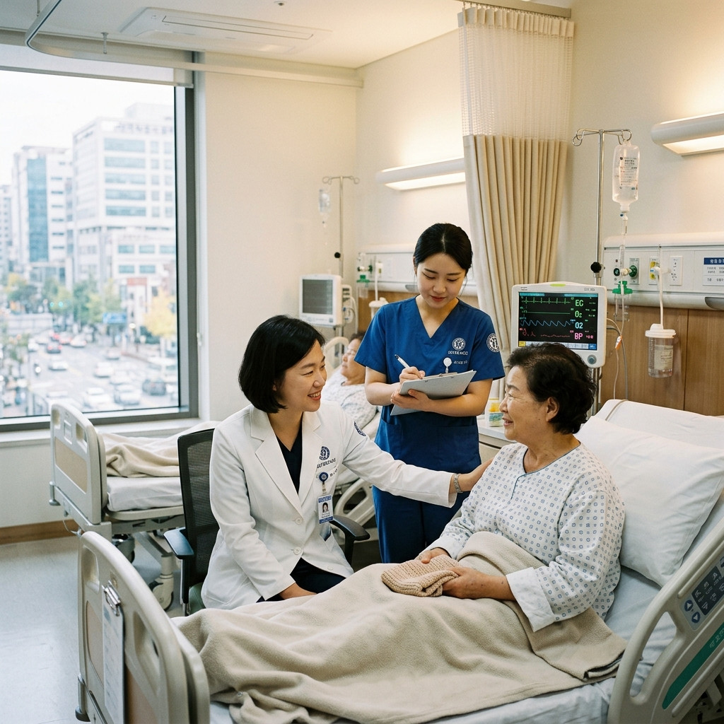
만인의 탑 현장 보도 실사
▲ 복부 영상의학 진단 및 혈액 췌장 효소 수치 정밀 판독 / 만인의 탑

2. 급성 췌장염 vs 만성 췌장염의 핵심 증상과 4대 비교 분석

급성 췌장염과 만성 췌장염은 발병 기전과 환자가 겪는 임상 양상에서 뚜렷한 차이를 보인다. 급성 췌장염의 가장 결정적인 징후는 갑작스럽게 찾아오는 명치와 윗배의 쥐어짜는 듯한 극심한 복통이다. 이 통증은 췌장이 후복막에 자리 잡고 있는 해부학적 특성상 등줄기와 날개뼈 사이, 허리 뒤쪽을 송곳으로 찌르는 듯한 강한 '방사통(Radiating pain)'을 필연적으로 동반한다. 똑바로 누우면 팽창한 췌장이 척추를 압박해 통증이 극에 달하지만, 무릎을 가슴으로 모으고 상체를 앞으로 웅크리는 새우잠 자세(기도 자세)를 취하면 일시적으로 통증이 완화되는 독특한 체위성 변화가 나타난다. 또한 구역질과 구토가 끊이지 않는데, 단순 위장염과 달리 구토를 쏟아내도 복통이 전혀 경감되지 않는다는 점이 감별 포인트다.

반면 만성 췌장염은 수년에 걸친 반복적인 염증 반응으로 췌장 실질 세포가 영구적으로 파괴되고 딱딱하게 굳어버린 비가역적 섬유화 및 석회화 질환이다. 급성기처럼 숨을 못 쉴 정도의 발작적 통증보다는 식후 30분에서 1시간 뒤 윗배가 더부룩하고 은근하게 쑤시는 둔통이 만성적으로 지속된다. 병이 진행될수록 지방을 소화하는 리파아제 분비가 바닥나면서 음식물 속 지방이 흡수되지 못해 변기 물에 기름이 둥둥 뜨고 심한 악취를 풍기는 '지방변(Steatorrhea)'과 만성 설사가 발생한다. 영양소 흡수 불량으로 수개월 만에 수 킬로그램의 체중이 급감하며, 인슐린을 분비하는 베타세포까지 소실되면서 기존 먹는 약으로는 조절이 극히 어려운 난치성 '췌장원성 3c형 당뇨병'이 동반된다.

구분 항목 급성 췌장염 (Acute) 만성 췌장염 (Chronic) 단순 위염 및 역류성 식도염
발병 기전 췌장 소화효소 급성 조기활성화 및 실질 자가소화 반복 염증에 의한 췌장 실질 비가역적 섬유화·석회화 위산 과다 및 위 점막 표재성 염증·식도 역류
통증 양상 쥐어짜는 명치 극통 및 척추 관통 등 방사통 식후 30분 뒤 반복되는 상복부 둔통 또는 무통증 식전 쓰림 또는 식후 타는 듯한 명치 흉통
체위별 반응 새우잠 자세(몸 숙임) 시 완화, 누우면 악화 체위 변화에 따른 통증 변화 미미 눕거나 상체를 숙이면 역류로 흉통 악화
핵심 동반 증상 구토 후에도 완화 안 됨, 미열·빈맥 동반 기름 둥둥 뜨는 악취 지방변, 체중 감소, 3c형 당뇨 제산제 복용 시 즉각 완화, 트림, 신물 올라옴
SPONSORED · ADVERTISEMENT

3. 공인 3대 진단 기준과 혈청 리파아제·아밀라아제 3배 상승의 의미

국제 표준 진단 기준인 '개정 애틀랜타 분류(Revised Atlanta Classification)'에 따르면, 급성 췌장염은 다음 세 가지 요건 중 두 가지 이상을 만족할 때 공식 확진 판정을 내린다. 첫째는 갑작스럽고 심하게 지속되는 명치 및 상복부 통증과 등 방사통이며, 둘째는 혈액 검사상 췌장 소화 효소인 혈청 아밀라아제(Amylase) 또는 리파아제(Lipase) 수치가 '정상 상한치(Upper Limit of Normal)의 3배 이상'으로 급상승한 경우다. 셋째는 조영증강 복부 전산화단층촬영(Enhanced Abdominal CT)이나 MRI, 복부 초음파 등 영상의학적 검사에서 췌장 부종이나 체액 저류, 췌장 주변 괴사 소견이 직접 확인되는 경우다.

임상에서 효소 수치가 정상 상한치의 3배 이상 치솟았다는 것은 단순한 자극을 넘어 췌장 선방세포막이 물리적으로 파괴되어 세포 속 효소가 전신 혈관으로 대량 누출되었음을 의미한다. 특히 혈청 리파아제는 아밀라아제에 비해 췌장 진단 특이도가 95% 이상으로 훨씬 높다. 아밀라아제는 타액선(침샘) 염증이나 신부전, 장폐색 등에서도 비특이적으로 상승할 수 있고 발병 3~5일 만에 빠르게 정상 수치로 떨어지는 한계가 있다. 반면 리파아제는 췌장에만 고유하게 존재하며 발병 후 8일에서 최대 14일까지 혈중에 고농도로 잔존하므로, 증상 발생 후 며칠이 지나 내원한 환자에서도 정확한 확진을 가능케 하는 핵심 지표다.

급성 췌장염 환자의 약 80%는 경증의 간질성 부종성 췌장염으로 며칠간의 치료로 회복되지만, 나머지 20%는 췌장 실질 조직이 썩어 들어가는 중증 괴사성 췌장염으로 악화되어 다발성 장기 부전으로 사망에 이를 수 있다. 따라서 즉각적인 3차 상급병원 응급실 이송이 요구되는 '치명적 경고 징후(Red Flags)'를 사전에 명확히 인지해야 한다. 첫째는 혈압이 수축기 90mmHg 이하로 급락하고 맥박수가 분당 120회를 웃도는 저혈압 패혈성 쇼크 증세다. 둘째는 염증 독소가 폐포 모세혈관을 공격해 호흡수가 분당 20회 이상으로 가빠지고 산소포화도가 떨어지는 급성 호흡곤란 증후군(ARDS)의 발생이다. 셋째는 배꼽 주변 피부가 푸르스름하게 착색되는 '쿨렌 징후(Cullen sign)'나 옆구리가 피멍이 든 것처럼 자줏빛으로 물드는 '그레이 터너 징후(Grey Turner sign)'다. 이 피부 징후들은 췌장의 자가소화로 파열된 혈관에서 흘러나온 피가 후복막강을 따라 피하 조직까지 번졌음을 뜻하는 중증 괴사성 출혈의 적신호다.

급성 췌장염 진단 직후 의학적으로 가장 먼저 취해지는 핵심 조치는 '절대 금식(NPO: Nil Per Os)'이다. 물 한 모금, 껌 한 조각이라도 입에 들어가는 순간 음식물을 감지한 신경계가 췌장에 소화 효소 분비 명령을 내려 자가소화를 폭발적으로 가속화하기 때문이다. 금식과 함께 췌장 모세혈관 혈류 공급을 유지하고 허혈성 괴사를 방어하기 위해 시간당 수백 밀리리터의 대량 정맥 수액(Lactated Ringer's solution)을 집중 주입하며, 극심한 통증은 마약성 진통제로 조절한다. 바터 팽대부를 막은 담석이 원인인 담석성 췌장염의 경우 응급 내시경적 역행성 담췌관 조영술(ERCP)을 시행해 담석을 즉각 제거해야 패혈증 진행을 막을 수 있다. 치료 후 재발 방지를 위해서는 단 한 잔의 술도 마시지 않는 완전한 단주가 평생 필수적이며, 고지방 식이를 엄격히 제한하고 혈중 중성지방 수치를 150mg/dL 이하로 철저히 관리해야 한다.

RECOMMENDED STORIES · SPONSORED
발행·편집인: 신승엽
Three Key Early Symptoms and Diagnostic Criteria of Pancreatitis: Severe Back-Radiating Epigastric Pain, Auto-Digestion Mechanism, and Acute vs. Chronic Differentiation
▲ Official Press Coverage & Contextual Archive / MANINTOP Global Newsroom
MANINTOP GLOBAL NEWSROOM · ENGLISH EDITION

Three Key Early Symptoms and Diagnostic Criteria of Pancreatitis: Severe Back-Radiating Epigastric Pain, Auto-Digestion Mechanism, and Acute vs. Chronic Differentiation

When excruciating epigastric pain strikes suddenly, radiating intensely through the spine to the back and forcing the patient into a fetal bending posture, it strongly signals pancreatitis—a severe medical emergency where the pancreas begins digesting its own tissue. Located deeply in the retroperitoneal cavity behind the stomach, the pancreas falls victim to fatal auto-digestion when gallstones or alcohol obstruction trap potent digestive enzymes. Grounded in clinical guidelines from the Korea Disease Control and Prevention Agency (KDCA) and pancreatic societies, we provide an in-depth analysis of the revised Atlanta diagnostic triad, chronic complications including steatorrhea and pancreatogenic diabetes, and emergency red flags demanding urgent intensive care.
📌 Key Takeaways
  • Verified factual reporting regarding Three Key Early Symptoms and Diagnostic Crite.
  • In-depth contextual and institutional analysis by MANINTOP Global Newsroom.
  • Official implementation schedule and verifiable public guidelines confirmed.

1. Retroperitoneal Anatomy and the Pathophysiology of Pancreatic Auto-Digestion

The pancreas is an elongated organ measuring approximately 15 centimeters, nestled horizontally in the deep retroperitoneal space directly behind the stomach and anterior to the spine and abdominal aorta. It functions simultaneously as an exocrine powerhouse—secreting 1.5 to 2 liters of digestive enzyme juice daily containing trypsinogen, amylase, and lipase—and an endocrine regulator secreting insulin and glucagon. Under healthy physiology, potent proteolytic enzymes are synthesized and transported as inactive proenzymes (trypsinogen) and are only cleaved into active trypsin by enterokinase upon reaching the duodenum, safeguarding pancreatic tissue.

However, when gallstones migrate from the gallbladder and lodge in the ampulla of Vater (the common junction of the bile and pancreatic ducts), or when chronic alcohol toxicity induces viscous intraductal protein plugs, pancreatic ductal pressure spikes precipitously. Trapped digestive enzymes undergo premature, abnormal intracellular activation within the pancreatic parenchyma. Active trypsin rapidly initiates devastating 'auto-digestion,' literally dissolving pancreatic acinar cells and adjacent vessels. The subsequent release of activated enzymes and cytokines into the retroperitoneal space triggers severe microvascular collapse, systemic inflammatory response syndrome (SIRS), and potentially fatal multiorgan failure.

2. Clinical Manifestations: Acute Attack vs. Chronic Pancreatic Insufficiency

Acute and chronic pancreatitis exhibit profoundly distinct pathophysiology and symptomatic profiles. Acute pancreatitis presents abruptly with excruciating, unrelenting pain concentrated in the epigastrium. Because of the retroperitoneal anatomy, this pain characteristically radiates straight through the back, scapulae, and lumbar region. Lying flat in the supine position exacerbates the agony as the inflamed organ compresses against the spine, whereas adopting a forward-bending 'prayer' or knee-to-chest posture alleviates retroperitoneal tension, offering temporary relief. Persistent nausea and vomiting accompany the episode; critically, unlike benign gastroenteritis, vomiting provides no relief whatsoever from the abdominal pain.

Conversely, chronic pancreatitis represents irreversible, progressive fibrosis and calcification resulting from recurrent inflammation over months or years. Instead of violent paroxysms, patients suffer from persistent or meal-induced dull aching in the upper abdomen. As lipase-producing acinar reserves diminish, unabsorbed dietary lipids are excreted directly in stool, producing classic 'steatorrhea'—foul-smelling, bulky, greasy feces that float in the toilet bowl. This malabsorption precipitates chronic diarrhea, fat-soluble vitamin deficiencies (A, D, E, K), and marked weight loss. Eventually, destruction of pancreatic islet beta cells leads to brittle, insulin-dependent pancreatogenic Type 3c diabetes.

3. Official Atlanta Diagnostic Criteria and the Significance of 3x Enzyme Elevation

Under the internationally recognized Revised Atlanta Classification, a definitive diagnosis of acute pancreatitis requires establishing at least two of the following three objective criteria. First, acute onset of persistent, severe epigastric pain often radiating to the back. Second, serum pancreatic enzyme activity—specifically lipase or amylase—at least three times above the laboratory upper limit of normal (ULN). Third, pathognomonic imaging findings of acute pancreatitis on contrast-enhanced abdominal computed tomography (CT), magnetic resonance imaging (MRI), or transabdominal ultrasound.

A serum enzyme concentration exceeding three times the upper normal threshold serves as a definitive clinical biomarker reflecting extensive acinar membrane disruption and massive enzymatic extravasation into circulation. Between the two biomarkers, serum lipase demonstrates superior clinical sensitivity and specificity exceeding 95%. While amylase can rise non-specifically in salivary gland disorders, bowel obstruction, or renal insufficiency and normalizes rapidly within 3 to 5 days, lipase is synthesized almost exclusively by the pancreas and remains elevated for 8 to 14 days, providing an indispensable diagnostic window for delayed presentations.

4. Emergency Red Flags Demanding Immediate ICU Admission and NPO Protocol

While approximately 80% of acute pancreatitis cases present as mild interstitial edematous inflammation that resolves with conservative care, the remaining 20% develop severe necrotizing pancreatitis associated with substantial morbidity and mortality. Recognizing critical 'red flags' is therefore vital for timely escalation to intensive care. These include hypotensive septic shock with systolic blood pressure falling below 90 mmHg and heart rate exceeding 120 beats per minute. A second warning sign is acute respiratory distress syndrome (ARDS), driven by systemic cytokine storms that compromise pulmonary alveolar capillaries. A third alarming physical indicator is cutaneous ecchymosis: Cullen's sign (bluish discoloration surrounding the umbilicus) or Grey Turner's sign (purplish bruising across the flank), signaling extensive retroperitoneal hemorrhage.

The cornerstone of acute management is absolute fasting (nil per os, NPO). Ingesting even a sip of water or chewing gum triggers neurohumoral pathways that stimulate pancreatic secretions, accelerating auto-digestion. Concurrently, aggressive intravenous hydration with balanced crystalloids (Lactated Ringer's) is initiated to sustain pancreatic microvascular perfusion and prevent ischemic necrosis, alongside tailored analgesia using opioid regimens. For gallstone pancreatitis with persistent ampullary obstruction, urgent endoscopic retrograde cholangiopancreatography (ERCP) with sphincterotomy is performed to extract impacted stones and prevent septic ascending cholangitis. Long-term recurrence prevention mandates lifelong, total abstinence from alcohol, rigorous adoption of a low-fat diet, and tight control of serum triglycerides below 150 mg/dL.

Category Acute Pancreatitis Chronic Pancreatitis Gastritis & Acid Reflux
Pathogenesis Acute early activation of pancreatic digestive enzymes and substantial autolysis Irreversible fibrosis and calcification of pancreatic tissue due to recurrent inflammation Excess gastric acid and superficial gastric mucosal inflammation, gastroesophageal reflux
Pain Patterns Cramping epigastric pain and radiating pain penetrating the spine Recurrent dull or painless upper abdominal discomfort 30 minutes after eating Burning sensation before meals or burning epigastric chest pain after meals
Positional Responses Relief in the fetal position (body bent forward), worsening when lying down Minimal changes in pain with position changes Worsening chest pain due to reflux when lying down or bending the upper body
Key Accompanying Symptoms No relief after vomiting, accompanied by low-grade fever and tachycardia Floating foul-smelling fatty stools, weight loss, type 3 diabetes Immediate relief upon taking antacids, belching, and regurgitation of bile
Publisher & Editor-in-Chief: Seung Youb Ssin, Ph.D.
Global Newsroom: MANINTOP GLOBAL NEWSROOM